
We segment the pelvis part with U-Net++, which is an improved version of U-Net. We use X-ray images of the pelvis to train our model, with 191 images as training data, and 33 images as validation data. Pixels in each image are labeled with two classes, where label 0 is background, and label 1 is pelvis. The model outputs a probability map with the size (N, H, W), where N is the number of classes; H and W are the height and width of input image. Each pixel represents the class prediction.
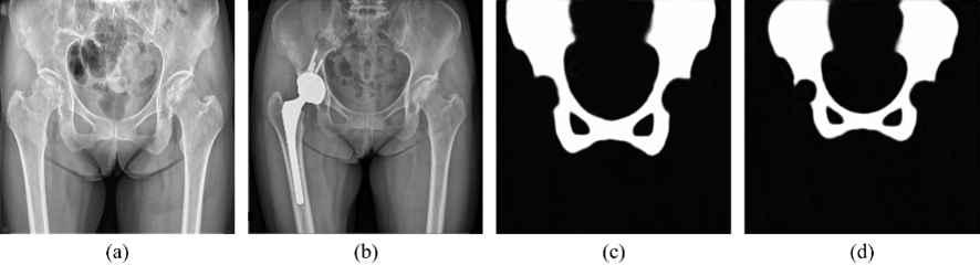 The probability map prediction result of our data. (a) X-ray image before surgery. (b) X-ray image after surgery. (c) Probability map prediction of pelvis before surgery. (d) After surgery. The pixel value represents the confidence score of pelvis.
Model created